
Article tag: BMN/B026| dog anatomy model| canine dissection model| veterinary anatomy teaching| half-muscle dog m|
How to teach canine anatomy with the BMN/B026 half-muscle dog model: what the exposed side shows, a lab plan, and where a model cannot replace dissection.
...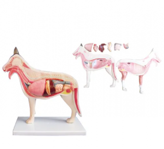
Veterinary anatomy has a practical problem that human anatomy does not: the specimen is expensive, ethically loaded and hard to store. A dog model that shows half the body in muscle and the other half opened to the organs solves the storage problem and much of the demonstration problem, at the cost of tactile realism.
The BMN/B026 is that model: a canine body with half-muscle dissection and the inner organs exposed for study. It is aimed at veterinary, zoology and biology teaching. This article sets out what the exposed side actually teaches, how to build a lab around it, and where the model must give way to a real specimen.
| Region | Structures visible | Teaching use |
|---|---|---|
| Body wall | Superficial muscles on the intact side | Palpation landmarks and muscle layers |
| Thoracic cavity | Heart and lungs in situ | Topographic relationships, not just organ names |
| Abdominal cavity | Digestive organs in position | Why organ placement explains clinical signs |
| Neck and head | Airway and proximal structures | Introduces the upper airway before dissection |
| Whole body | Standing posture on a base | Orientation and surface anatomy |
The value is topographic. Students often learn organs as a list — liver, stomach, spleen — without the spatial relationships that make a clinical examination meaningful. A model that keeps organs in position teaches the relationship; a chart does not.
Step five is what makes the model useful in a veterinary programme rather than a biology display: it prepares the student for the dissection lab, where specimen time is limited and expensive.
Physical models are strong for spatial learning but do not substitute for cadaveric dissection where haptic learning is the objective [1]. A programme that uses the model to replace dissection will produce students who recognise structures in a photograph but hesitate with a scalpel.
Quotation and specification. Current pricing, dimensions and delivery for the BMN/B026 are available from cprmodel@adaanatomy.com.
Q1. What does the BMN/B026 show? A dog with half the body in muscle and the other half opened to expose the internal organs, for canine anatomy study.
Q2. Is it life-size? It is a teaching model on a base; ask the supplier for the exact dimensions, since display scale varies by model.
Q3. Who uses it? Veterinary, zoology and biology programmes teaching canine structure and topography.
Q4. Can it replace dissection? No. It teaches spatial relationships; tissue feel and variation require a specimen.
Q5. Are the organs removable? Confirm with the supplier — models differ in which organs are fixed and which can be taken out.
Q6. How should it be cleaned and stored? Dust with a soft brush or damp cloth, avoid solvents, and store upright on its base away from direct sun.
Product page: Teaching Canine Anatomy with the BMN/B026 Half-Muscle Dog Model — specification, packing data and a quotation request form.
For a quotation on the BMN/B026, or to ask which organs are detachable, write to cprmodel@adaanatomy.com with your programme level and intake.