
Article tag: BIX-A1074 lung segment model| transparent lung model| bronchial tree model| 2X lung anatomy model| lung |
Transparent 2X lung model BIX-A1074 — segment structure and bronchial tree visible through the shell; built for anatomy, surgery and internal medicine classes.
...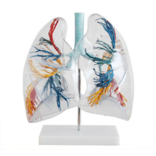
Short answer: BIX-A1074 is a two-times enlarged lung model with a transparent outer shell. The page states that it shows the segments — eight parts of the left lung and ten of the right — and that the distribution of the bronchial tree can be observed through the transparent shell. The lung outside is transparent plastic and the tracheobronchial tree is plastic. It is a visual teaching aid for medical colleges, thoracic surgery and internal medicine.
Key takeaways
| Item | Published detail |
|---|---|
| Model | BIX-A1074 |
| Category | Medical Series → Lung Models |
| Scale | 2 times magnification |
| Segments | 8 parts of the left lung; 10 parts of the right lung |
| Materials | Transparent plastic outer lung; plastic tracheal and bronchial tree |
| Intended use | Visual teaching in medical colleges: anatomy, thoracic surgery, internal medicine |
| Package size / weight | Not published |
| Price | Not published — quotation required |
Lung anatomy is taught by segments because surgery is done by segments. A model that shows lobes only cannot support a conversation about a segmentectomy; one that shows eight left and ten right segments can. The published count is the standard division — eight on the left, ten on the right — so the model maps directly onto the textbook diagram students are examined on.
The transparent shell adds the second half of the lesson: the bronchial tree. Students can follow a bronchus from the carina into a specific segment without the model being cut open. That is the relationship that makes segment anatomy clinically useful, because the bronchus is the road map to the segment.
| Aspect | Assessment |
|---|---|
| Segment visibility | Strong. Eight left and ten right segments are stated, matching standard anatomy. |
| Bronchial tree | Strong. Visible through the transparent shell, so the airway-to-segment link can be traced. |
| Size for teaching | 2X magnification works on a bench for a group of eight to ten. |
| Pathology | None. No tumour, consolidation, emphysema or effusion is represented. |
| Palpation and auscultation | None. This is a visual model; pair it with a torso or an auscultation trainer for those skills. |
| Durability | Transparent polymers can cloud; confirm the cleaning agent with the supplier. |
Segment anatomy is one of the areas where three-dimensional visualisation has measurable value. A 2025 study of 3D visualisation and printing in teaching lung segment anatomy reported improved spatial understanding compared with two-dimensional resources [1]. A 2026 evaluation of a low-cost simulation tool for diagnostic bronchoscopy training showed that a physical airway model can carry procedural teaching, not only anatomy [2]. A 2025 review of 3D printing in medicine describes the broader pattern: physical models bridge imaging and the classroom, but they must be designed for a specific teaching task [3].
The limitation follows from that last point. BIX-A1074 is designed for visual anatomy, so it should be judged on segment clarity and bronchial tree legibility — not on whether it feels like lung tissue.
| Not stated on the page | Question to ask |
|---|---|
| Overall dimensions and weight | Needed for shelf space and freight. |
| Whether the model can be opened | Affects cleaning and whether the bronchial tree can be replaced. |
| Colour coding of segments | Colour makes segment identification faster in a large class. |
| Stand or base included | A 2X model needs stable support for demonstration. |
| Price and lead time | Quotation item. |
Email cprmodel@adaanatomy.com with the model BIX-A1074 and your class size, and we will send the dimensions, packing data and quotation together.
Q1. What is BIX-A1074?
A 2X transparent lung model that shows the segments and the bronchial tree for anatomy teaching.
Q2. How many segments does it show?
Eight parts of the left lung and ten parts of the right lung, as stated on the product page.
Q3. Can you see the bronchi?
Yes. The page states that the distribution of the bronchial tree can be observed through the transparent shell.
Q4. Is it suitable for auscultation practice?
No. It is a visual anatomy model with no lung sounds or palpation features.
Q5. What is it made of?
Transparent plastic for the outer lung and plastic for the tracheal and bronchial tree.
Q6. How do I get a price?
Email cprmodel@adaanatomy.com with the model (BIX-A1074), quantity and delivery city, and ask for the dimensions and packing data in the same message.
Write to cprmodel@adaanatomy.com for the BIX-A1074 specification sheet and a quotation. If you teach a full respiratory module, ask in the same email which lung and larynx models share the same scale, so the demonstration set is consistent on the bench.