
Article tag: ADA-436| uterus model| life-size uterus anatomy| cervix anatomy model| midwifery teaching model|
ADA-436 versus a sectioned pelvis model: what a life-size coronal uterus shows, what a pelvis adds, and which one your course should buy first.
...
Two purchases get confused constantly: a life-size uterus model and a sectioned female pelvis. Both are sold as "pelvic anatomy", both sit on a bench, and both are bought by the same programmes. They teach different things, and buying the wrong one is the most common equipment mistake in midwifery and gynaecology teaching.
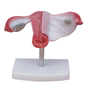
The ADA-436 is a life-size uterus with a coronal section. The listing specifies that it shows the uterus, the cervix divided into supravaginal and vaginal portions, the eight ligaments of the uterus and the fallopian tubes. That list is the clue to what it is for.
| Feature | ADA-436 uterus model | Sectioned pelvis model |
|---|---|---|
| Scope | Uterus, cervix, ligaments, tubes | Pelvis, bladder, rectum, uterus in situ |
| Section | Coronal | Usually sagittal or median |
| Scale | Life-size organ | Pelvis girdle, organ relationships |
| Strongest lesson | Cervical and ligament anatomy | Spatial relationships and surgical approach |
| Typical user | Midwifery, gynaecology, nursing | Surgery, radiology, obstetrics |
| Weakness | No surrounding structures | Organ detail often simplified |
The cervix in two parts. The supravaginal and vaginal portions of the cervix are a genuine exam favourite and a genuine clinical distinction — the supravaginal portion sits above the vaginal attachment, the vaginal portion projects into the vagina. A coronal section shows the relationship in a single view, which is exactly how it should be taught.
The eight ligaments. The uterus is held by a set of ligaments whose names students memorise without understanding direction. A model that shows all eight lets an instructor ask a better question: which ligament, if shortened, changes the position of the fundus?
The tubes and their path. The fallopian tube is not a straight line; the model shows the curve and the fimbrial end, which matters for teaching fertility and ectopic pregnancy.
The uterus does not sit alone. A sectioned pelvis shows it between bladder and rectum, with the pouch of Douglas behind and the pubic symphysis in front. That relationship is what a clinician needs when interpreting an ultrasound or planning an approach.
Three-dimensional visualisation of the third stage of labour has been studied in midwifery education, and qualitative findings showed that students working with a three-dimensional representation were better able to apply their knowledge to practice [1]. The lesson for buyers: the value is in the spatial representation, not in the number of parts. A uterus model provides that representation for the organ; a pelvis provides it for the cavity.
A common and defensible sequence:
Buying the pelvis first and the uterus never is the mistake. Students then know where the uterus is but not what it is made of, and they struggle with cervical assessment, IUCD placement and postpartum examination.
Retention research is blunt on this point: nursing students' gross anatomy knowledge declines measurably after the foundational course [2]. A life-size organ model that lives in the skills lab gives students a way to re-check anatomy while practising a procedure, which is the kind of revisiting that retention requires.
State these limits; students who expect a model to answer every question stop trusting it when it cannot.
Quotation and specification. Pricing, dimensions and delivery for the ADA-436 are available from cprmodel@adaanatomy.com.
Q1. Is the ADA-436 life-size? Yes. It is specified as a natural, life-size uterus model.
Q2. What does the coronal section show? The uterine cavity and walls, the cervix with its supravaginal and vaginal portions, the eight uterine ligaments and the fallopian tubes.
Q3. Does it include ovaries? The listing specifies the uterus, cervix, ligaments and tubes. Confirm ovary inclusion with the supplier if your course requires the adnexa.
Q4. Can it be used for IUCD insertion training? No. It is an anatomy model. Insertion training needs a dedicated task trainer with a simulated cervix and cavity.
Q5. Which should a midwifery programme buy first? The uterus model, then a sectioned pelvis. Organ anatomy precedes spatial relationships in most curricula.
Q6. Does it show pregnancy changes? No. It represents the non-pregnant uterus.
Product page: ADA-436 Uterus Model vs Sectioned Pelvis: What Each One Actually Shows — specification, packing data and a quotation request form.
For a quotation on the ADA-436, write to cprmodel@adaanatomy.com with your programme and intake.