
Article tag: BIX-A1112| lymphatic system model| lymphoid model| lymphatic anatomy teaching| anatomy lab equipment|
BIX-A1112 buyer's guide: what a lymphatic wall model must show, how to check organ coverage, and where a static model stops and physiology begins.
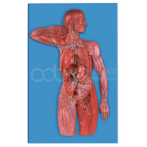
...
The lymphatic system is the part of anatomy most often taught in a hurry at the end of a term — and the part students retain least well. That is not only a student problem. The system is diffuse, it overlaps with the venous return, and it has almost no landmarks that a beginner can hold on to.
A wall model such as the BIX-A1112 addresses the first difficulty by putting the whole system on one surface. Whether it earns a place in your lab depends on what it shows and how you teach with it.
| Item | Detail | Why it matters |
|---|---|---|
| Subject | Composition and structural characteristics of the lymphatic system | Confirms whole-system scope, not a single organ |
| Size | 76 × 49 × 8.5 cm | Wall-mounted or easel display; check wall space |
| Format | Single display panel | Whole-system view in one glance |
| Use | Anatomy and immunology teaching | Supports both structural and functional framing |
The 76 × 49 cm format is a deliberate teaching choice: large enough to read from the back of a tutorial room, flat enough to hang. Confirm whether the model is supplied with a mounting rail or easel, because a panel that must be propped against a wall will be used less.
Check these six items before buying any lymphatic model:
A model that shows nodes but not directionality teaches a map without traffic. Students will be able to label the stations and still fail to explain why an infection in the hand produces axillary tenderness.
Lymphatic anatomy is a network problem: many small vessels, many nodes, one destination. Textbooks compress it into a schematic, and schematics lose the spatial sense of how much territory drains where.
Physical models of dense anatomical relationships have a documented advantage over three-dimensional computer models and textbooks for learning spatial anatomy [1]. The lymphatic system is a good test case, because the relationships are the content.
Retention is the second problem. Studies of nursing students show that gross anatomy knowledge declines after the foundational course unless it is revisited in context [2]. A wall model in the corridor or skills lab serves exactly that function: it makes revision incidental rather than scheduled.
Be explicit about these limits, or students will treat the panel as a complete account of the system.
Three-dimensional printing has been used to build task-specific models for lymph node dissection education, and a randomised trial found the printed model improved learning for lateral pelvic node dissection [3]. That is a reminder that the best lymphatic teaching tool depends on the task: a wall panel for overview, a printed or sectional model for procedure.
Quotation and specification. Pricing, mounting options and delivery for the BIX-A1112 are available from cprmodel@adaanatomy.com.
Q1. What size is the BIX-A1112? 76 × 49 × 8.5 cm, a wall-panel format intended for group teaching.
Q2. Does it show the thoracic duct? The model is specified to show the composition and structural characteristics of the lymphatic system; confirm duct labelling on the unit, since this is the single most important structure for teaching drainage.
Q3. Is it a 3D relief model or a flat panel? The listing gives a thickness of 8.5 cm, which suggests a relief model rather than a printed chart. Confirm before ordering if relief is essential.
Q4. Can it be used for immunology teaching? It supports organ-level immunology. Cellular and histological immunology need separate resources.
Q5. Is it suitable for nursing programmes? Yes, particularly for revision of node groups and drainage routes used in assessment.
Q6. What should we pair it with? A lymph node section model for histology, and a venous system model for the re-entry point.
Product page: BIX-A1112 Lymphatic System Model: A Procurement Guide for Anatomy Labs — specification, packing data and a quotation request form.
For a quotation on the BIX-A1112, or to confirm mounting hardware, write to cprmodel@adaanatomy.com.