
Article tag: BIX-A1106| palm anatomy model| palmar layers model| hand anatomy teaching| palmar fascia|
Palmar anatomy on the BIX-A1106 model: the three palmar layers, why the fascia matters clinically, and how to teach safe corridors without a cadaver.
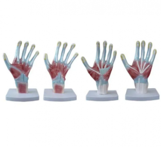
...
The palm is a small space with a lot of consequence. A centimetre in the wrong direction changes a cut from a minor wound into a hand that cannot oppose a thumb. Students learn the palm badly because it is taught as a list of structures rather than as a stack of layers, and layers are exactly what a physical model shows best.
The BIX-A1106 is a palm model that displays the anatomical levels of the palm from superficial to deep. This article sets out what those levels are, why they matter clinically, and how to teach them so the knowledge survives the exam.
| Level | Structures | Clinical relevance |
|---|---|---|
| 1. Skin and fat | Palmar skin, fibrous fat pads | Skin is thick and fixed; lacerations gape little but hide depth |
| 2. Palmar fascia | Palmar aponeurosis, fibrous septa | Dupuytren contracture arises here |
| 3. Tendon layer | Flexor tendons in synovial sheaths | Sheath infection spreads along the digit |
| 4. Neurovascular layer | Median and ulnar nerves, superficial arch | The "safe corridor" question lives at this level |
| 5. Deep layer | Deep palmar arch, interossei, metacarpals | Deep space infections and hardware planning |
Teaching the palm as five levels rather than forty structures cuts the memorisation load and explains why the same cut can be trivial or catastrophic.
The palmar aponeurosis is not a wrapping; it is a structural element with pathological behaviour. Dupuytren disease — thickening and contracture of the palmar fascia — is common enough that most hand clinics see it weekly, and its treatment carries real complication risk, including nerve injury and recurrence [1].
That single fact changes how the fascia should be taught. It is not a grey sheet in a diagram; it is the tissue that decides whether a patient can lay the hand flat. A model that shows the aponeurosis as a distinct layer, with the septa running to the skin, makes the disease mechanism legible before students ever see a contracture.
The classic teaching is that incisions in the palm should avoid the thenar crease and the hypothenar area, and should follow the creases. The reason is the neurovascular layer beneath.
Use the model for a three-step exercise:
Students who perform step 3 once remember the corridor. Students who read it in a table often do not.
The palmaris longus tendon is a convenient landmark — and it is absent in a substantial minority of people. Clinical examination studies across populations have reported agenesis rates that vary widely by group, which is precisely why it cannot be assumed [2]. In a model with a fixed anatomy, that variation disappears.
Say this out loud in class. A model teaches one normal; a patient may present another. The model's job is to give a reference strong enough that a variation is noticed, not to define the only possible arrangement.
Spatial relationships in the hand are dense, and physical models have been shown to outperform three-dimensional computer models and textbooks for learning such relationships [3]. The palm is an extreme case: five layers in a few centimetres, with structures crossing at different angles.
A screen lets a student rotate the hand; a model lets them lift a layer and see what is under it. That difference matters when the lesson is about depth and risk.
Quotation and specification. Pricing, packing and delivery for the BIX-A1106 are available from cprmodel@adaanatomy.com.
Q1. What does the BIX-A1106 show? It shows the anatomical levels of the palm from superficial to deep, allowing layer-by-layer study of palmar structures.
Q2. Is it a full hand or only the palm? The model is described as a palm model. Confirm with the supplier whether the digits are included if your course covers digital sheaths.
Q3. Can it be used for surgical training? It is an anatomy teaching model, not a procedural simulator. It supports preoperative planning teaching, not instrument practice.
Q4. Does it show nerves and vessels? The layered design is intended to expose deeper structures as superficial layers are removed; confirm the specific structures on the unit before purchase.
Q5. How many units per carton? Ten sets per carton, 74 × 43 × 29 cm, 12 kg.
Q6. What should we buy with it? A hand skeleton for bone detail, and a flexor tendon sheath model if infection spread is in the syllabus.
Product page: Palmar Anatomy on the BIX-A1106 Model: Layers, Tendons and Safe Corridors — specification, packing data and a quotation request form.
For a quotation on the BIX-A1106, write to cprmodel@adaanatomy.com with the course it will support.