
Article tag: BIX-A1054| liver anatomical model| hepatic vascular anatomy| portal vein model| bile duct teaching m|
Teaching liver vascular anatomy with the BIX-A1054: colour coding, portal versus biliary routes, and the segment logic students must not skip.
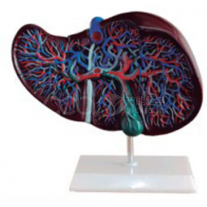
...
The liver is the organ where students first meet two incoming blood supplies and one outgoing duct, and where a single colour mistake destroys the whole lesson. A model that separates the routes by colour is not a luxury; it is the only way to teach the arrangement in one session.
The BIX-A1054 represents the complete vascular network of the liver in different colours: the portal blood vessel, and the intrahepatic and extrahepatic bile ducts. This article explains how to use the colour coding to teach the portal triad, and where the model's logic ends.
| Item | Detail |
|---|---|
| Model | BIX-A1054 liver anatomical model |
| Vascular structures | Complete hepatic vascular network |
| Colour coding | Separate colours for portal vessels and biliary routes |
| Biliary detail | Intrahepatic and extrahepatic bile ducts |
| Format | Whole-organ teaching model |
| Intended use | Gross anatomy, hepatobiliary anatomy teaching |
Everything in hepatobiliary teaching rests on students knowing that the portal triad carries three things in two directions: portal vein in, hepatic artery in, bile duct out. Colour coding makes that visible, and visibility is what most students lack when they first meet a liver specimen.
Run the session in this order:
That fourth step is where models usually fail. A liver model that shows inflow and biliary drainage but not hepatic venous drainage teaches two-thirds of the circulation, and students will draw the missing third incorrectly in exams.
Once the routes are clear, the model becomes a map for segments. Cadaveric and imaging studies of hepatic segmentation show why surgeons care: the segments are defined by vascular territories, not by surface landmarks, and the surface gives almost no clue to what lies beneath [3].
Three-dimensional visualisation and printed models have been shown to improve students' understanding of hepatic segments compared with two-dimensional images alone [1][2]. A physical model with colour-coded vessels is the low-cost version of the same intervention: it lets students see that a segment is a vascular unit.
Quotation and specification. Current pricing, packing and delivery for the BIX-A1054 are available from cprmodel@adaanatomy.com.
Q1. What does the BIX-A1054 show? The complete hepatic vascular network in different colours, including the portal blood vessel and the intrahepatic and extrahepatic bile ducts.
Q2. Does it show the hepatic vein? Confirm with the supplier which vessels are colour-coded; the specified structures are the portal vessel and the biliary routes. Teach hepatic venous drainage explicitly if the model omits it.
Q3. Is it suitable for nursing courses? Yes for gross anatomy, though nursing syllabi usually need less vascular detail than surgical courses.
Q4. Can it be used to teach liver segments? It supports segment teaching because the vessels are visible, but you will need a segmentation chart or imaging to define the eight segments.
Q5. What is it made of? PVC-type teaching material with a painted colour code; confirm the exact material and finish with the supplier.
Q6. What should we order with it? A liver segment chart or 3D-printed segment model, and a biliary system model if the course covers obstruction and jaundice.
Product page: Teaching Hepatic Vascular Anatomy with the BIX-A1054 Liver Model — specification, packing data and a quotation request form.
For a quotation on the BIX-A1054, or to ask which vessels are colour-coded, write to cprmodel@adaanatomy.com.