
Article tag: BMN/B045| frog anatomy model| amphibian dissection model| 3X frog model| biology teaching|
How to run a frog practical with the BMN/B045 3X model: pre-lab mapping, organ removal, comparison with a real specimen, and the limits of a model.
...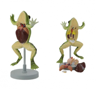
Frog dissection survives in biology curricula because it teaches something rare: a whole vertebrate, small enough to see at once. The problem is supply, ethics and time. The BMN/B045 — a 3X life-size frog with the ventral surface opened and removable components — changes the arithmetic of that practical without removing the animal from the syllabus.
This guide is for the instructor who wants the model to do real work rather than sit at the front of the lab as a demonstration piece.
| Feature | Detail | Classroom consequence |
|---|---|---|
| Scale | 3X life size | Every structure visible from the back row |
| Orientation | Ventral surface opened | Direct comparison with a ventral dissection |
| Components | Removable organs | Removal and replacement drills |
| Use | Amphibian anatomy teaching | Pre-lab rehearsal, post-lab revision |
Three times life size is the key number. At that scale a student can see the heart, the liver lobes and the gut coil without leaning over a tray, which means the instructor can teach to the whole group instead of repeating the same demonstration eight times.
Give students the model before they meet the specimen. A 15-minute pre-lab with the model changes what they do with the dissection:
Students who arrive with a map dissect faster and damage less tissue. The model is not replacing the specimen; it is making the specimen legible.
Place one model at each bench and keep it open while students work on the specimen. The most common failure in a frog dissection is not squeamishness — it is loss of orientation after the first cut. The model restores orientation in seconds.
Use it for three specific checks:
This is the step most courses skip and the one that pays for the model. Ask students to list three things the model got right and three things the specimen showed that the model did not. Typical answers:
| Model advantage | Specimen advantage |
|---|---|
| Structures are clean and separated | Real tissue planes and texture |
| No smell, no preservative protocol | Colour and vascularity are real |
| Organs can be removed repeatedly | Variation between individuals |
Then make the point explicit: a model teaches arrangement; a specimen teaches variation. A course that uses both produces students who can handle a frog they have never seen.
The published evidence supports this pairing. A systematic review of controlled studies on alternatives to animal use in education found that alternatives can be effective, but did not find them uniformly superior to animal-based teaching [1]. The defensible position is not replacement but sequencing: model first, specimen second, and say so in the course outline.
Where a programme reduces animal numbers, the standard argument is threefold: fewer animals, repeatable practice, and no scheduling constraints. The model supports all three. It also removes the preservative handling protocol from the pre-lab, which shortens setup time and reduces the ventilation requirement in the lab.
What it does not remove is the ethical discussion. Students still need to understand why the animal was used and what alternatives exist; the model is a good prompt for that conversation, not a way to avoid it.
Quotation and specification. Pricing, component list and delivery for the BMN/B045 are available from cprmodel@adaanatomy.com.
Q1. Is the BMN/B045 life-size? No. It is a 3X life-size model, enlarged so that structures are visible to a class.
Q2. Can it replace a real dissection? It can replace the demonstration, but not the tissue-handling experience. Use it before and after the specimen session [1].
Q3. How many organs are removable? The listing specifies removable components but does not give a count. Confirm the parts list with the supplier.
Q4. Is it suitable for high-school biology? Yes. At 3X scale it suits large classes and avoids preservative handling for younger students.
Q5. Does it show the ventral or dorsal surface? The ventral part is opened to allow study of the internal anatomy.
Q6. What should we buy with it? A frog skeleton for the skeletal comparison, and a hand lens set for small structures.
Product page: Running a Frog Dissection Practical with the BMN/B045 3X Model — specification, packing data and a quotation request form.
For a quotation on the BMN/B045, or to request the component list, write to cprmodel@adaanatomy.com.