
Article tag: BIX-A1110| neural system model| spinal nerve model| peripheral nerve teaching| nervous system anatom|
Teaching spinal nerve distribution on the BIX-A1110: how to read the board, where students go wrong, and the three limits of a flat model.
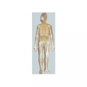
...
Students can name the brachial plexus and still fail to say which nerve a needle passes near. The gap is spatial: the plexus is taught as a diagram, and diagrams hide depth. A full-length nervous system board puts the cord, the plexus and the terminal nerves back into one continuous map.
The BIX-A1110 shows the central nervous system, the spinal cord and the peripheral spinal nerves, including the spinal nerves running from the central nervous system to the limbs — the radial, ulnar and median nerves among them. The model measures 83 × 30 × 5 cm, so it presents the whole axis in one view. This article sets out how to use it and where its limits begin.
| Item | Detail |
|---|---|
| Model | BIX-A1110 neural system model |
| Dimensions | 83 × 30 × 5 cm |
| Structures shown | Central nervous system, spinal cord, peripheral spinal nerves |
| Named limb nerves | Radial, ulnar, median and other terminal branches |
| Format | Full-axis board model |
| Intended use | Neuroanatomy and peripheral nerve teaching |
Most courses teach the central nervous system and the peripheral nervous system in different weeks, from different figures, in different proportions. Students then assemble them mentally, and that assembly is where errors appear.
A single 83 cm board forces continuity. The spinal cord is not a stub that ends at the foramen magnum — it runs the length of the model, with roots exiting at each level and the plexus forming where the limb nerves gather. When a student can trace one continuous line from cord to fingertip, the "peripheral" nervous system stops being a separate subject.
Neuroanatomy teaching research supports the same conclusion in a different format: three-dimensional and cross-sectional representations improve learning of spatially complex structure, and students consistently perform better when the representation preserves the relationships rather than listing the parts [1]. A flat board preserves relationships along one axis, which is why it works for the cord-to-limb question and fails for anything requiring depth.
Step 1 — levels, not names (10 min). Ask students to count the spinal nerve pairs shown and mark where the cord thickens. The cervical and lumbar enlargements are visible, and the reason for them is limb innervation, not anatomy trivia.
Step 2 — follow one nerve (15 min). Give each pair of students one named nerve — radial, ulnar, median — and ask them to trace it from cord to hand, naming each region it crosses. Groups then compare routes. The comparison is the lesson: three nerves, three different paths, one plexus.
Step 3 — clinical application (15 min). Ask what a lesion at three different points would cost the patient: at the root, in the plexus, at the wrist. Students who can answer using the board have understood distribution rather than memorised labels.
Spatial ability predicts performance in anatomy, and physical models help most where mental rotation is weakest [3]. That argues for handling, not demonstration: pass the board around, or mount it low enough that students can point.
Quotation and specification. Current pricing, packing and delivery for the BIX-A1110 are available from cprmodel@adaanatomy.com.
Q1. What does the BIX-A1110 show? The central nervous system, the spinal cord and the peripheral spinal nerves, including the spinal nerves running to the limbs.
Q2. Which named nerves are visible? The radial, ulnar and median nerves are among the terminal branches shown, together with the spinal nerves along the axis.
Q3. How big is it? 83 × 30 × 5 cm.
Q4. Is it suitable for physiotherapy or nursing courses? Yes. It suits any course that teaches peripheral nerve distribution and the consequences of nerve injury.
Q5. Can it show cranial nerves? Not in detail. Use a brain or head model for cranial nerve anatomy.
Q6. What should we order with it? A limb cross-section model for depth, and a brain model for the central structures the board shows only in outline.
Product page: Mapping Spinal Nerves on the BIX-A1110 Neural System Model — specification, packing data and a quotation request form.
For a quotation on the BIX-A1110, or to ask about mounting options and the labelled key, write to cprmodel@adaanatomy.com.